


Vitamin D and Bone Density: What Your DEXA Scan Reveals About Vitamin D Deficiency
Half of all adults in the United Kingdom have lower-than-optimal vitamin D levels, yet most have no idea their bones may be paying the price. Vitamin D is the gatekeeper of calcium absorption: without enough of it, your body cannot move calcium from your diet into the bone tissue where it is needed most. Over months and years, that silent shortfall thins the internal scaffolding of your skeleton, raising your risk of fractures, osteoporosis, and chronic pain.
A blood test can tell you whether your vitamin D level is low. A bone density DEXA scan tells you whether that deficiency has already affected your skeleton. Together, they give a complete picture that neither test provides alone.
Quick Answer: Vitamin D regulates calcium absorption. When levels drop below 25 nmol/L, bone mineral density can decline even if calcium intake is adequate. A DEXA scan measures that bone loss directly, identifying damage that a blood test cannot detect. The NHS recommends 10 micrograms of vitamin D daily for every adult, rising to 20 micrograms for those with osteoporosis.
Why Vitamin D Matters for Bone Density
Vitamin D is a fat-soluble hormone that controls how much calcium your intestines absorb from food. When your vitamin D level is sufficient (above 50 nmol/L in blood), your gut absorbs roughly 30 to 40 percent of dietary calcium. When levels fall below 25 nmol/L, that figure drops to around 10 to 15 percent, according to the Royal Osteoporosis Society.
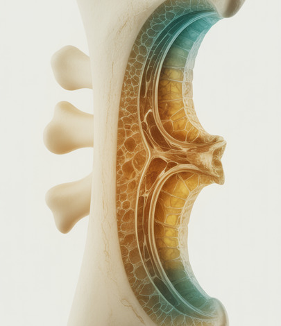
Your body responds by pulling calcium out of your bones to maintain blood calcium levels for nerve and muscle function. This process, called bone resorption, weakens the trabecular (spongy) bone inside your hip, spine, and wrist. If it continues unchecked, the result is reduced bone mineral density and, eventually, osteoporosis.
Unlike many nutrients, vitamin D cannot be obtained in sufficient quantities from diet alone. The primary source is sunlight on exposed skin, and in the UK your skin can only produce vitamin D between April and September. During autumn and winter, stored vitamin D may not last, which is why public health guidelines recommend supplementation for all adults during those months.
How Common Is Vitamin D Deficiency in the UK?
The scale of vitamin D deficiency in the United Kingdom is far larger than most people realise. Data from the UK Biobank and national surveys show that around one in five adults has levels below 25 nmol/L (clinical deficiency), while roughly half have levels below the optimal threshold of 50 nmol/L.
Certain groups face significantly higher risk. People with darker skin (those of African, African-Caribbean, or South Asian heritage) produce vitamin D more slowly through UV exposure. In the UK Biobank study, 57 percent of participants with Asian ancestry were deficient during winter and spring, compared with 18 percent of White European participants. Older adults, people who spend most of their time indoors, those who cover most of their skin, and individuals living in northern regions of the UK are also at elevated risk.
The challenge is that vitamin D deficiency rarely produces obvious symptoms in its early stages. You can be losing bone mineral density for years before a fracture, a fall, or a routine scan reveals the damage.
What a Blood Test Shows and What It Misses
A serum 25-hydroxyvitamin D blood test is the standard way to check your vitamin D status. NHS guidelines classify results as follows: below 25 nmol/L is deficient, 25 to 50 nmol/L is insufficient, and above 50 nmol/L is sufficient for most adults.
A blood test is useful because it tells you what is happening right now. It can confirm deficiency, guide supplementation doses, and track whether treatment is working. However, it cannot tell you what has already happened to your bones. Vitamin D levels fluctuate with the seasons, with diet, and with sun exposure. A single reading in June might look reassuring even if you spent years with low levels through the winter months.
This is the gap that a DEXA scan fills. A DEXA scan measures bone mineral density at the hip and spine with precision better than one percent. It records the cumulative effect of every factor that has acted on your skeleton, including years of vitamin D insufficiency that no single blood test can reconstruct.
How a DEXA Scan Reveals the Damage Vitamin D Deficiency Causes
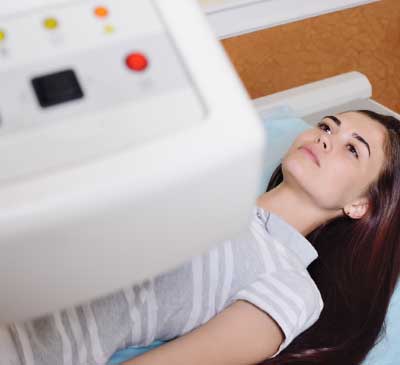
DEXA (dual-energy X-ray absorptiometry) works by passing two low-dose X-ray beams through your bones. The scanner measures how much energy each beam loses, and from that calculates bone mineral density in grams per square centimetre. The result is expressed as a T-score (compared with a healthy young adult) and a Z-score (compared with someone of your age and sex).
A T-score above -1.0 is considered normal. Between -1.0 and -2.5 indicates osteopenia (reduced bone density). Below -2.5 indicates osteoporosis. The scan takes around ten minutes, uses very low radiation (less than a day of natural background exposure), and requires no preparation.
For someone with low vitamin D, the DEXA result shows the real-world consequence of that deficiency. It can reveal thinning at the lumbar spine or femoral neck that blood work alone would never detect. This is clinically important because fracture risk doubles with each standard deviation decline in bone mineral density, according to guidelines from the National Osteoporosis Guideline Group (NOGG).
Combining a vitamin D blood test with a body composition DEXA scan gives both cause and effect: the blood test explains why bone loss may be occurring, and the DEXA scan shows whether it has.
Who Should Consider a DEXA Scan for Vitamin D-Related Bone Loss?
Not everyone with low vitamin D needs a DEXA scan immediately, but several groups benefit from combining both tests. You should consider a scan if you have had a confirmed vitamin D deficiency (below 25 nmol/L) for more than twelve months, if you have a family history of osteoporosis or fragility fractures, or if you are a postmenopausal woman (oestrogen decline accelerates bone loss independently of vitamin D).
Other candidates include adults over 50 with risk factors such as low body weight, smoking history, or long-term use of corticosteroids (which both suppress vitamin D metabolism and directly weaken bone). People who have already fractured a bone from a minor fall or bump should be scanned regardless of vitamin D status, as the fracture itself suggests bone density may already be compromised.
If you fall into any of these categories, a DEXA scan at our Harley Street clinic provides a precise baseline. Repeat scans at intervals recommended by your clinician (typically every two to three years) can track whether supplementation and lifestyle changes are protecting or rebuilding bone.
Protecting Your Bones: Vitamin D, Calcium, and What the Evidence Shows
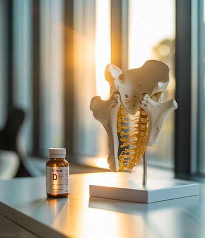
The NHS recommends that every adult in the UK takes 10 micrograms (400 IU) of vitamin D daily, with supplementation especially important from October through March when sunlight is insufficient. For those with osteoporosis or taking bisphosphonate medication, the dose rises to 20 micrograms (800 IU), as recommended by the Royal Osteoporosis Society.
Evidence on supplementation is nuanced. A meta-analysis published in The Lancet Diabetes and Endocrinology found that vitamin D supplements alone do not significantly improve bone density for adults with sufficient baseline levels. However, in adults who are genuinely deficient (below 30 nmol/L), supplementation does improve bone mineral density and may reduce fracture risk, particularly when combined with adequate calcium intake of 700 to 1200 mg per day.
Weight-bearing exercise (walking, jogging, dancing, resistance training) stimulates bone formation independently of vitamin D. The combination of correcting a deficiency, ensuring adequate calcium, and regular weight-bearing activity gives your skeleton the best chance of maintaining or recovering density. A DEXA scan before and after these interventions provides measurable confirmation that your strategy is working.
Frequently Asked Questions
Can vitamin D deficiency cause osteoporosis?
Prolonged vitamin D deficiency contributes to osteoporosis by reducing calcium absorption and triggering bone resorption. It is one of several modifiable risk factors, alongside low physical activity, smoking, and inadequate calcium intake.
How much vitamin D should I take for bone health?
The NHS recommends 10 micrograms (400 IU) daily for all UK adults. If you have osteoporosis or are on bone-protective medication, your doctor may recommend 20 micrograms (800 IU). Do not exceed 100 micrograms (4,000 IU) daily without medical supervision.
Will a DEXA scan show if I have vitamin D deficiency?
A DEXA scan does not measure vitamin D levels directly. It measures bone mineral density, which reveals the skeletal consequences of deficiency. Pairing a DEXA scan with a vitamin D blood test gives the most complete picture.
How often should I have a DEXA scan if I am vitamin D deficient?
For adults at moderate risk, a scan every two to three years is typical. High-risk individuals (those with confirmed osteoporosis or on treatment) may benefit from scans every one to two years to track response to therapy.
Is it too late to improve bone density if I have been deficient for years?
It is rarely too late. Correcting a deficiency, adding weight-bearing exercise, and ensuring adequate calcium can slow further bone loss and, in some cases, modestly increase bone mineral density. A DEXA scan establishes your starting point so progress can be tracked objectively.
Book Your DEXA Bone Density Scan at Harley Street
If you are concerned about vitamin D deficiency and its impact on your bone health, a DEXA scan provides the clinical evidence you need. At DEXA London, our Harley Street clinic offers precise bone mineral density measurement in a scan that takes around ten minutes with no preparation required.
Your results are reported by Dr Emil Gadimali and include your T-score, Z-score, and site-specific density readings at the hip and lumbar spine. Combined with a vitamin D blood test from your GP, these results give you and your clinician a clear, data-driven foundation for protecting your bones.
To book your scan, call 0207 637 8227 or visit our clinic at 86 Harley Street, London.
Half of all adults in the United Kingdom have lower-than-optimal vitamin D levels, yet most have no idea their bones may be paying the price. Vitamin D is the gatekeeper of calcium absorption: without enough of it, your body cannot move calcium from your diet into the bone tissue where it is needed most. Over months and years, that silent shortfall thins the internal scaffolding of your skeleton, raising your risk of fractures, osteoporosis, and chronic pain.
A blood test can tell you whether your vitamin D level is low. A bone density DEXA scan tells you whether that deficiency has already affected your skeleton. Together, they give a complete picture that neither test provides alone.
Quick Answer: Vitamin D regulates calcium absorption. When levels drop below 25 nmol/L, bone mineral density can decline even if calcium intake is adequate. A DEXA scan measures that bone loss directly, identifying damage that a blood test cannot detect. The NHS recommends 10 micrograms of vitamin D daily for every adult, rising to 20 micrograms for those with osteoporosis.
Why Vitamin D Matters for Bone Density
Vitamin D is a fat-soluble hormone that controls how much calcium your intestines absorb from food. When your vitamin D level is sufficient (above 50 nmol/L in blood), your gut absorbs roughly 30 to 40 percent of dietary calcium. When levels fall below 25 nmol/L, that figure drops to around 10 to 15 percent, according to the Royal Osteoporosis Society.
Your body responds by pulling calcium out of your bones to maintain blood calcium levels for nerve and muscle function. This process, called bone resorption, weakens the trabecular (spongy) bone inside your hip, spine, and wrist. If it continues unchecked, the result is reduced bone mineral density and, eventually, osteoporosis.
Unlike many nutrients, vitamin D cannot be obtained in sufficient quantities from diet alone. The primary source is sunlight on exposed skin, and in the UK your skin can only produce vitamin D between April and September. During autumn and winter, stored vitamin D may not last, which is why public health guidelines recommend supplementation for all adults during those months.
How Common Is Vitamin D Deficiency in the UK?
The scale of vitamin D deficiency in the United Kingdom is far larger than most people realise. Data from the UK Biobank and national surveys show that around one in five adults has levels below 25 nmol/L (clinical deficiency), while roughly half have levels below the optimal threshold of 50 nmol/L.
Certain groups face significantly higher risk. People with darker skin (those of African, African-Caribbean, or South Asian heritage) produce vitamin D more slowly through UV exposure. In the UK Biobank study, 57 percent of participants with Asian ancestry were deficient during winter and spring, compared with 18 percent of White European participants. Older adults, people who spend most of their time indoors, those who cover most of their skin, and individuals living in northern regions of the UK are also at elevated risk.
The challenge is that vitamin D deficiency rarely produces obvious symptoms in its early stages. You can be losing bone mineral density for years before a fracture, a fall, or a routine scan reveals the damage.
What a Blood Test Shows and What It Misses
A serum 25-hydroxyvitamin D blood test is the standard way to check your vitamin D status. NHS guidelines classify results as follows: below 25 nmol/L is deficient, 25 to 50 nmol/L is insufficient, and above 50 nmol/L is sufficient for most adults.
A blood test is useful because it tells you what is happening right now. It can confirm deficiency, guide supplementation doses, and track whether treatment is working. However, it cannot tell you what has already happened to your bones. Vitamin D levels fluctuate with the seasons, with diet, and with sun exposure. A single reading in June might look reassuring even if you spent years with low levels through the winter months.
This is the gap that a DEXA scan fills. A DEXA scan measures bone mineral density at the hip and spine with precision better than one percent. It records the cumulative effect of every factor that has acted on your skeleton, including years of vitamin D insufficiency that no single blood test can reconstruct.
How a DEXA Scan Reveals the Damage Vitamin D Deficiency Causes
DEXA (dual-energy X-ray absorptiometry) works by passing two low-dose X-ray beams through your bones. The scanner measures how much energy each beam loses, and from that calculates bone mineral density in grams per square centimetre. The result is expressed as a T-score (compared with a healthy young adult) and a Z-score (compared with someone of your age and sex).
A T-score above -1.0 is considered normal. Between -1.0 and -2.5 indicates osteopenia (reduced bone density). Below -2.5 indicates osteoporosis. The scan takes around ten minutes, uses very low radiation (less than a day of natural background exposure), and requires no preparation.
For someone with low vitamin D, the DEXA result shows the real-world consequence of that deficiency. It can reveal thinning at the lumbar spine or femoral neck that blood work alone would never detect. This is clinically important because fracture risk doubles with each standard deviation decline in bone mineral density, according to guidelines from the National Osteoporosis Guideline Group (NOGG).
Combining a vitamin D blood test with a body composition DEXA scan gives both cause and effect: the blood test explains why bone loss may be occurring, and the DEXA scan shows whether it has.
Who Should Consider a DEXA Scan for Vitamin D-Related Bone Loss?
Not everyone with low vitamin D needs a DEXA scan immediately, but several groups benefit from combining both tests. You should consider a scan if you have had a confirmed vitamin D deficiency (below 25 nmol/L) for more than twelve months, if you have a family history of osteoporosis or fragility fractures, or if you are a postmenopausal woman (oestrogen decline accelerates bone loss independently of vitamin D). For more on recognising the warning signs, see our guide to early signs of osteoporosis and when women should get a bone density scan.
Other candidates include adults over 50 with risk factors such as low body weight, smoking history, or long-term use of corticosteroids (which both suppress vitamin D metabolism and directly weaken bone). People who have already fractured a bone from a minor fall or bump should be scanned regardless of vitamin D status, as the fracture itself suggests bone density may already be compromised.
If you fall into any of these categories, a DEXA scan at our Harley Street clinic provides a precise baseline. Repeat scans at intervals recommended by your clinician (typically every two to three years) can track whether supplementation and lifestyle changes are protecting or rebuilding bone.
Protecting Your Bones: Vitamin D, Calcium, and What the Evidence Shows
The NHS recommends that every adult in the UK takes 10 micrograms (400 IU) of vitamin D daily, with supplementation especially important from October through March when sunlight is insufficient. For those with osteoporosis or taking bisphosphonate medication, the dose rises to 20 micrograms (800 IU), as recommended by the Royal Osteoporosis Society.
Evidence on supplementation is nuanced. A meta-analysis published in The Lancet Diabetes and Endocrinology found that vitamin D supplements alone do not significantly improve bone density for adults with sufficient baseline levels. However, in adults who are genuinely deficient (below 30 nmol/L), supplementation does improve bone mineral density and may reduce fracture risk, particularly when combined with adequate calcium intake of 700 to 1200 mg per day.
Weight-bearing exercise (walking, jogging, dancing, resistance training) stimulates bone formation independently of vitamin D. The combination of correcting a deficiency, ensuring adequate calcium, and regular weight-bearing activity gives your skeleton the best chance of maintaining or recovering density. A DEXA scan before and after these interventions provides measurable confirmation that your strategy is working.
Frequently Asked Questions
Can vitamin D deficiency cause osteoporosis?
Prolonged vitamin D deficiency contributes to osteoporosis by reducing calcium absorption and triggering bone resorption. It is one of several modifiable risk factors, alongside low physical activity, smoking, and inadequate calcium intake.
How much vitamin D should I take for bone health?
The NHS recommends 10 micrograms (400 IU) daily for all UK adults. If you have osteoporosis or are on bone-protective medication, your doctor may recommend 20 micrograms (800 IU). Do not exceed 100 micrograms (4,000 IU) daily without medical supervision.
Will a DEXA scan show if I have vitamin D deficiency?
A DEXA scan does not measure vitamin D levels directly. It measures bone mineral density, which reveals the skeletal consequences of deficiency. Pairing a DEXA scan with a vitamin D blood test gives the most complete picture.
How often should I have a DEXA scan if I am vitamin D deficient?
For adults at moderate risk, a scan every two to three years is typical. High-risk individuals (those with confirmed osteoporosis or on treatment) may benefit from scans every one to two years to track response to therapy.
Is it too late to improve bone density if I have been deficient for years?
It is rarely too late. Correcting a deficiency, adding weight-bearing exercise, and ensuring adequate calcium can slow further bone loss and, in some cases, modestly increase bone mineral density. A DEXA scan establishes your starting point so progress can be tracked objectively.
Book Your DEXA Bone Density Scan at Harley Street
If you are concerned about vitamin D deficiency and its impact on your bone health, a DEXA scan provides the clinical evidence you need. At DEXA London, our Harley Street clinic offers precise bone mineral density measurement in a scan that takes around ten minutes with no preparation required.
Your results are reported by Dr Emil Gadimali and include your T-score, Z-score, and site-specific density readings at the hip and lumbar spine. Combined with a vitamin D blood test from your GP, these results give you and your clinician a clear, data-driven foundation for protecting your bones.
To book your scan, call 0207 637 8227 or visit our clinic at 86 Harley Street, London.