


Type 2 Diabetes and Body Composition: What Your DEXA Scan Reveals About Diabetes Risk
Most people associate type 2 diabetes risk with sugar intake or family history. Fewer realise that body composition plays a central and measurable role in determining how likely someone is to develop the condition. The ratio of fat to lean tissue, where that fat sits, and how much muscle mass you carry all influence insulin sensitivity in ways that standard weight checks cannot detect.
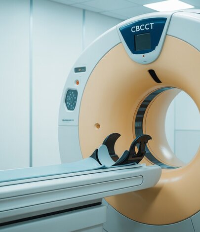
A DEXA (dual-energy X-ray absorptiometry) scan provides a detailed, region-by-region breakdown of your body composition. It quantifies visceral fat, subcutaneous fat, lean mass, and bone density with clinical precision. For anyone concerned about metabolic health or diabetes risk, this information is far more useful than a number on the scales.
Quick Answer: A body composition DEXA scan measures visceral fat, lean muscle mass, and fat distribution with clinical accuracy. These metrics are stronger predictors of type 2 diabetes risk than BMI or body weight alone. Elevated visceral fat and low muscle mass both independently increase insulin resistance, and a DEXA scan can detect these patterns before symptoms appear.
How Body Composition Affects Type 2 Diabetes Risk
Type 2 diabetes develops when the body becomes resistant to insulin or when the pancreas can no longer produce enough insulin to manage blood glucose levels. While genetics and lifestyle both contribute, research consistently shows that body composition is one of the strongest modifiable risk factors.
According to Diabetes UK, approximately 90% of people diagnosed with type 2 diabetes are living with overweight or obesity. However, the overall amount of body fat tells only part of the story. Two people with identical body weights can have very different metabolic profiles depending on their fat distribution and muscle mass.
A 2021 study published in Diabetologia found that individuals with high visceral fat but normal BMI had significantly elevated fasting glucose and HbA1c levels compared to those with similar BMI but lower visceral fat. This pattern, sometimes referred to as “metabolically obese, normal weight,” highlights why composition matters more than total mass.
Why BMI Misses the Full Picture
Body mass index (BMI) divides weight by height squared. It does not distinguish between fat and muscle, nor does it account for where fat is stored. The NHS uses BMI as a screening tool, and for population-level research it remains useful. At the individual level, however, it frequently misclassifies metabolic risk.
A person with significant muscle mass may register as overweight on the BMI scale while having excellent metabolic markers. Conversely, someone within the “healthy” BMI range may carry dangerous amounts of visceral fat around their organs. A study in the Annals of Internal Medicine found that adults with normal BMI but central obesity had the highest mortality risk of any weight category.
For assessing diabetes risk specifically, BMI cannot tell you how much visceral adipose tissue you carry, whether you are losing muscle as you age, or how your fat distribution compares to clinical thresholds. A DEXA scan can answer all three of these questions in a single appointment.
What a DEXA Scan Measures That Matters for Diabetes
A DEXA scan breaks the body down into three compartments: bone mineral content, lean soft tissue (primarily muscle), and fat mass. It maps each compartment across distinct body regions, including the trunk, arms, legs, and the android (abdominal) zone.
The metrics most relevant to diabetes risk include:
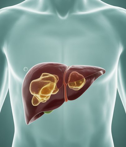
- Visceral adipose tissue (VAT): DEXA estimates the fat stored around and between your abdominal organs. High VAT is directly linked to insulin resistance, elevated triglycerides, and increased inflammatory markers.
- Android-to-gynoid fat ratio (A/G ratio): This compares abdominal fat to hip and thigh fat. A higher ratio indicates a more metabolically risky fat distribution pattern.
- Appendicular lean mass index (ALMI): This measures lean tissue in the arms and legs relative to height. Low ALMI is associated with impaired glucose disposal and increased diabetes risk, particularly in older adults.
- Total body fat percentage: While less specific than regional measures, total fat percentage provides useful context alongside the metrics above.
Together, these measurements create a far more complete picture of metabolic risk than any single number can provide.
Visceral Fat and Insulin Resistance
Visceral fat is the single most important body composition metric for diabetes risk assessment. Unlike subcutaneous fat (the fat you can pinch beneath the skin), visceral fat is metabolically active. It releases inflammatory cytokines, free fatty acids, and hormones that directly interfere with insulin signalling.
Research published in The Lancet Diabetes & Endocrinology has shown that visceral fat volume is a stronger predictor of incident type 2 diabetes than total body fat, waist circumference, or BMI. The relationship is dose-dependent: the more visceral fat you carry, the greater your insulin resistance tends to be.
A DEXA scan quantifies visceral fat in grams and assigns a risk category. For our patients at DEXA London, we have found that this number often provides the clearest early warning of metabolic trouble, sometimes years before blood glucose levels cross the diagnostic threshold for prediabetes. We explored this relationship in depth in our guide to what your visceral fat DEXA results reveal about metabolic health.
Lean Muscle Mass and Blood Sugar Control
While much of the diabetes conversation focuses on fat, muscle mass deserves equal attention. Skeletal muscle is the body’s largest glucose sink. After a meal, muscle tissue is responsible for absorbing roughly 80% of glucose from the bloodstream in an insulin-mediated process.
When muscle mass is low, the body has fewer sites available for glucose uptake, which can lead to elevated blood sugar levels even in the absence of excess fat. A 2022 meta-analysis in The Journal of Clinical Endocrinology & Metabolism found that low skeletal muscle mass was independently associated with a 50% increased risk of developing type 2 diabetes, after adjusting for age, sex, and body fat.
This is particularly relevant for older adults, people following calorie-restricted diets without adequate protein or resistance training, and individuals on GLP-1 receptor agonist medications where lean mass preservation requires monitoring. A DEXA scan can track lean mass over time and flag meaningful losses before they become clinically significant.
Tracking Your Progress with Repeat DEXA Scans
A single DEXA scan provides a valuable baseline. Repeat scans, typically at 6 to 12 month intervals, reveal how your body composition is changing in response to lifestyle modifications, exercise programmes, dietary changes, or medical interventions.
For people working to reduce their diabetes risk, tracking changes in visceral fat and lean mass over time provides objective evidence of progress. The scales might not move, but a DEXA scan can show that you have lost two kilograms of visceral fat and gained a kilogram of muscle. That kind of recomposition represents a meaningful reduction in metabolic risk, even if body weight stays the same.
The NHS recommends that people at risk of type 2 diabetes aim for a 5 to 10% reduction in body weight. A DEXA scan adds nuance to this target by showing whether weight loss is coming from fat (desirable) or muscle (undesirable). This distinction is critical for long-term metabolic health and helps guide more effective interventions.
Frequently Asked Questions
Can a DEXA scan diagnose type 2 diabetes?
No. A DEXA scan measures body composition, not blood glucose or HbA1c levels. However, the metrics it provides, particularly visceral fat and lean mass, are strong predictors of diabetes risk and can prompt earlier investigation with blood tests.
I have a normal BMI. Could I still be at risk?
Yes. Research shows that people with normal BMI but high visceral fat or low muscle mass can have elevated diabetes risk. A DEXA scan can identify this pattern, which BMI alone cannot detect.
How often should I have a DEXA scan to monitor my risk?
For most people, a scan every 6 to 12 months is appropriate. This interval is long enough for measurable changes to occur in body composition while being frequent enough to track trends and adjust your approach if needed.
Will losing visceral fat reduce my diabetes risk?
The evidence is clear that reducing visceral fat improves insulin sensitivity. The NHS Diabetes Prevention Programme cites visceral fat reduction as a key outcome associated with lower progression rates from prediabetes to type 2 diabetes.
Is a DEXA scan safe?
DEXA uses a very low dose of radiation, roughly equivalent to one or two days of natural background exposure. It is considered safe for routine clinical use and takes approximately 10 to 15 minutes.
Book Your DEXA Scan at DEXA London
Understanding your body composition is one of the most practical steps you can take to assess and manage your type 2 diabetes risk. At DEXA London, our clinic on Harley Street provides clinical-grade DEXA body composition scans with a detailed report covering visceral fat, lean mass, fat distribution, and bone density.
Every scan is reviewed by Dr Emil Gadimali, and your results come with a clear explanation of what each metric means for your metabolic health. Whether you are looking for a baseline assessment or tracking changes over time, our team is here to help you make sense of the numbers.
To book your scan, call us on 0207 637 8227 or visit our body composition scan page to learn more about what the appointment involves.
Weight management next step
If your DEXA results point to elevated visceral fat or metabolic risk factors associated with type 2 diabetes, a supervised weight-loss programme may help reduce your risk. CutKilo, the sister service to DEXA London, offers doctor-led Mounjaro (tirzepatide) treatment supervised by Dr Emil Gadimali. Start the CutKilo questionnaire to see if you are suitable.