

Chronic Kidney Disease and Body Composition: What Your DEXA Scan Reveals About CKD-Related Muscle and Bone Changes
Chronic kidney disease (CKD) affects more than 10% of the global population, and its impact reaches far beyond the kidneys themselves. As kidney function declines, the body undergoes significant changes in muscle mass, fat distribution, and bone mineral density. These shifts often progress silently, meaning patients may not realise the extent of the damage until a fracture occurs or mobility declines sharply.
A body composition DEXA scan provides a detailed, clinical-grade assessment of all three compartments: fat mass, lean mass, and bone mineral density. For people living with CKD, this information can be transformative, offering an early window into complications that blood tests alone may miss.
Quick answer: CKD accelerates muscle wasting (with sarcopenia prevalence reaching 25% across all CKD stages and over 50% in advanced disease), disrupts bone mineral metabolism through a condition called CKD-MBD, and alters fat distribution in ways that increase cardiovascular risk. A DEXA scan is one of the most reliable ways to track these changes and guide treatment decisions.
How Chronic Kidney Disease Affects Your Body Composition
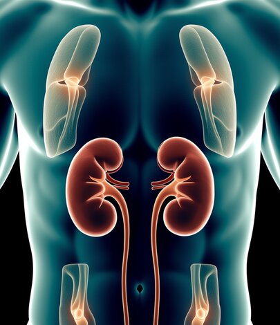
The kidneys do far more than filter waste from the blood. They regulate calcium and phosphate balance, activate vitamin D, produce erythropoietin for red blood cell production, and help control acid-base balance throughout the body. When kidney function drops below normal, each of these processes is disrupted, and the consequences show up directly in body composition.
CKD triggers a cascade of metabolic disturbances. Metabolic acidosis breaks down muscle protein. Elevated parathyroid hormone (PTH) leaches calcium from bones. Chronic inflammation and reduced physical activity accelerate fat accumulation around the organs. According to a 2024 systematic review published in the Journal of Cachexia, Sarcopenia and Muscle, CKD patients experience simultaneous losses in lean mass and bone density while often gaining visceral fat, a combination that dramatically increases cardiovascular and fracture risk.
What makes this particularly challenging is that standard clinical markers like body weight and BMI can remain stable even as muscle is replaced by fat. A patient may weigh the same as they did a year ago, yet their body composition DEXA scan could reveal a significant shift from lean tissue to fat tissue. This is why body composition assessment, rather than simple weight monitoring, is increasingly recognised as essential in CKD management.
CKD-Related Muscle Loss: Why Sarcopenia Is So Common in Kidney Disease
Sarcopenia, the progressive loss of skeletal muscle mass and function, is one of the most clinically significant complications of CKD. A 2024 global meta-analysis found that approximately 25% of all CKD patients meet the diagnostic criteria for sarcopenia, with prevalence rising sharply in advanced stages. Among patients on dialysis, the figure exceeds 50% in some studies.
Several mechanisms drive muscle wasting in CKD. Metabolic acidosis, which develops as the kidneys lose their ability to excrete acid, directly stimulates muscle protein breakdown through the ubiquitin-proteasome pathway. Chronic systemic inflammation, elevated levels of myostatin (a protein that inhibits muscle growth), insulin resistance, and reduced physical activity all compound the problem. The dialysis procedure itself has been shown to stimulate protein degradation and suppress protein synthesis, making muscle preservation even harder for patients receiving renal replacement therapy.
The consequences extend well beyond physical appearance. CKD-related sarcopenia is independently associated with higher hospitalisation rates, increased mortality, poorer quality of life, and greater difficulty performing daily activities. Research published in Frontiers in Nutrition (2025) confirms that the relationship between CKD and sarcopenia is bidirectional: kidney disease accelerates muscle loss, and reduced muscle mass may itself worsen kidney function.
A DEXA scan quantifies appendicular lean mass (the muscle in your arms and legs) with precision, making it possible to detect sarcopenia at an early stage, well before grip strength or walking speed decline noticeably. For CKD patients, this early detection window is critical. Our guide to age-related muscle loss and what your DEXA scan reveals after 40 explores how sarcopenia develops and why early measurement matters.
Bone Mineral Disorder in CKD: What Happens to Your Skeleton
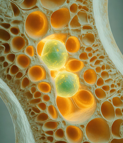
CKD-Mineral and Bone Disorder (CKD-MBD) is a systemic condition that encompasses abnormalities in calcium, phosphorus, PTH, and vitamin D metabolism, along with changes in bone turnover, mineralisation, and volume. It is almost universal in advanced CKD and represents one of the most important reasons to monitor bone density in kidney patients.
As kidney function declines, the kidneys lose their ability to convert vitamin D into its active form (calcitriol). Without adequate calcitriol, calcium absorption from the gut drops, blood calcium levels fall, and the parathyroid glands respond by producing more PTH. This secondary hyperparathyroidism accelerates bone resorption, pulling calcium out of the skeleton and weakening it progressively. According to the Kidney Disease: Improving Global Outcomes (KDIGO) guidelines, patients with CKD stages 3 to 5 face a 2 to 14-fold increase in fracture risk compared to the general population.
A 2025 study published in Frontiers in Medicine using advanced 3D-DXA technology found that patients with CKD stages G3b to G5 had significantly lower cortical bone parameters compared to those with earlier-stage disease, and that most cortical bone measurements correlated directly with glomerular filtration rate (GFR). In other words, the worse the kidney function, the thinner and weaker the cortical bone.
A bone density DEXA scan measures bone mineral density at the hip and lumbar spine, the sites most vulnerable to fracture. The KDIGO 2017 guideline update (reaffirmed in 2023) recommends that clinicians consider BMD testing in CKD patients when the results would influence treatment decisions, particularly for those at risk of osteoporosis or fragility fractures.
Why a DEXA Scan Is Essential If You Have CKD
Standard CKD monitoring includes blood tests for creatinine, eGFR, calcium, phosphate, PTH, and vitamin D. While these markers are essential, they cannot tell you how much muscle you have, where your fat is stored, or how strong your bones actually are. A DEXA scan fills that gap by providing direct, quantified measurements of all three compartments in a single appointment lasting around 15 minutes.
For CKD patients specifically, a DEXA scan offers several clinical advantages. It detects sarcopenia before functional decline becomes obvious, by measuring appendicular lean mass and comparing it to established reference ranges. It quantifies bone mineral density to assess fracture risk, which is especially important given the complex bone pathology in CKD-MBD. It measures visceral adipose tissue (VAT), which is strongly associated with cardiovascular mortality in CKD. And it provides a reproducible baseline for tracking the effects of treatment over time.
DEXA is particularly valuable because it uses a very low radiation dose (less than a day of natural background radiation) and is non-invasive. For patients who are already managing multiple appointments and interventions, a quick and painless scan that delivers this level of clinical insight is an efficient use of time. To understand more about the technology, see our page on how DEXA scanning works.
What Your DEXA Results Mean When You Have Kidney Disease
Interpreting DEXA results in the context of CKD requires some additional nuance compared to the general population. Here is what each key measurement tells you.
Appendicular Lean Mass (ALM): This measures the lean tissue in your arms and legs and is the primary marker used to diagnose sarcopenia. In CKD patients, ALM may decline even when total body weight is stable, because muscle is being replaced by fat or fluid. Serial DEXA scans (typically every 6 to 12 months) can track whether your lean mass is holding steady, declining, or responding to exercise and nutritional interventions.
Bone Mineral Density (BMD): Your T-score compares your bone density to that of a healthy young adult. In CKD, a low T-score may reflect osteoporosis, renal osteodystrophy, or a combination of both. While DEXA cannot distinguish between different types of bone disease (a bone biopsy is needed for that), it remains the most practical and widely available tool for assessing fracture risk. The KDIGO guidelines support its use in CKD stages 3a through 5D when the results will guide treatment.
Visceral Adipose Tissue (VAT): CKD patients often accumulate visceral fat even without significant overall weight gain. Elevated VAT is associated with inflammation, insulin resistance, and cardiovascular events. Your DEXA report will show your VAT area in square centimetres, and your clinician can use this alongside your kidney function markers to assess metabolic risk.
Total Body Fat Percentage: In CKD, the combination of rising fat mass and falling lean mass can produce a normal BMI while masking a high-risk body composition profile. This pattern, sometimes called sarcopenic obesity, carries a worse prognosis than either condition alone.
How to Protect Your Muscle and Bone Health With CKD
While CKD cannot be reversed in most cases, the body composition changes it causes can be slowed, stabilised, or partially reversed with the right interventions. A DEXA scan provides the baseline data needed to target treatment effectively.
Resistance exercise is one of the most evidence-supported strategies for preserving muscle mass in CKD. Even modest, supervised strength training programmes have been shown to improve lean mass, physical function, and quality of life in patients across all CKD stages, including those on dialysis. Your nephrologist or physiotherapist can help design a programme that accounts for any cardiovascular or musculoskeletal limitations.
Nutritional optimisation is equally important. CKD patients need careful protein management: enough to prevent muscle wasting, but not so much that it overloads the kidneys. Current evidence from the Indian Journal of Nephrology (2024) suggests that dietary counselling focused on protein quality and timing, combined with correction of metabolic acidosis, can meaningfully reduce sarcopenia risk.
Vitamin D supplementation may help address the deficiency that drives secondary hyperparathyroidism and bone loss, though the form and dose must be guided by your renal team. Active vitamin D analogues (such as calcitriol or alfacalcidol) are often needed because the kidneys cannot perform the final activation step.
Managing weight and visceral fat through a combination of dietary changes and physical activity can reduce the inflammatory and metabolic burden on the kidneys. Research published in Kidney International (2025) highlights the bidirectional relationship between obesity and CKD, with weight management identified as a modifiable factor that can slow disease progression.
Frequently Asked Questions
Is a DEXA scan safe for people with kidney disease?
Yes. A DEXA scan uses an extremely low dose of radiation, significantly less than a standard chest X-ray and equivalent to less than a day of natural background exposure. It does not require any contrast dye or injection, which makes it safe for people with impaired kidney function. There are no known contraindications specific to CKD.
How often should CKD patients have a DEXA scan?
For most CKD patients, a DEXA scan every 12 months is appropriate. If you are starting a new treatment (such as a bone-protective medication, a structured exercise programme, or a weight management plan), your clinician may recommend scanning at 6-month intervals to track early changes. The KDIGO guidelines support BMD testing when results will inform clinical decisions.
Can a DEXA scan detect CKD-MBD specifically?
A DEXA scan measures bone mineral density, which is one component of CKD-MBD. It cannot distinguish between different types of renal bone disease (such as high-turnover versus low-turnover bone disease), as that requires a bone biopsy. However, it remains the most practical, non-invasive, and widely available tool for assessing fracture risk in CKD patients, and the KDIGO Controversies Conference (2023) reaffirmed its clinical utility.
Will my DEXA results be different because I have CKD?
CKD can affect DEXA results in several ways. Fluid retention (common in advanced CKD) may slightly inflate lean mass measurements. Vascular or joint calcifications can artificially raise BMD readings at certain sites. An experienced clinician will take these factors into account when interpreting your scan. At DEXA London, your results are reviewed by Dr Emil Gadimali, who considers your full clinical context alongside the scan data.
Should I tell my nephrologist about my DEXA scan results?
Absolutely. Sharing your DEXA results with your renal team gives them valuable information about your bone and muscle health that blood tests alone cannot provide. This can influence decisions about vitamin D supplementation, phosphate binders, exercise referrals, and whether bone-protective medications are appropriate.
Book Your DEXA Scan at Our Harley Street Clinic
If you are living with chronic kidney disease and want a clear, detailed picture of how CKD is affecting your muscle mass, bone density, and body fat distribution, a DEXA scan at DEXA London can provide the answers. Our clinic at 86 Harley Street offers body composition and bone density scans in a single appointment, with results reviewed by Dr Emil Gadimali.
To book your scan or discuss whether a DEXA assessment is right for your situation, call us on 0207 637 8227 or book online through our website. Bring your most recent blood results to your appointment so we can interpret your scan in the fullest clinical context.
Weight management and kidney health
If your DEXA results show elevated visceral fat or a body composition profile that may be placing additional strain on your kidneys, supervised weight management can be an important part of protecting your renal function. CutKilo, the sister service to DEXA London, offers doctor-led Mounjaro treatment from Dr Emil Gadimali. Start the CutKilo questionnaire to see if you are suitable.