
Fatty Liver Disease and Body Composition: What Your DEXA Scan Reveals About Liver Health Risk
Key Findings
You step on the scales, and the number looks fine. Your BMI falls within the normal range. Yet an ultrasound has just revealed fat deposits in your liver. How is that possible?
Fatty liver disease, now formally known as metabolic dysfunction-associated steatotic liver disease (MASLD), affects roughly one in five UK adults. Many of them have no idea. The condition develops silently, driven less by total body weight and more by where fat accumulates inside the body. That distinction matters because a body composition DEXA scan can measure the exact regional fat distribution that standard scales and BMI calculations miss entirely.
Understanding your trunk fat percentage, your visceral adipose tissue levels, and the ratio of abdominal to peripheral fat gives both you and your clinician a far clearer picture of metabolic liver risk than body weight alone.
Quick answer: A DEXA scan measures trunk fat percentage and visceral adipose tissue, the two strongest predictors of fatty liver disease (MASLD). Research shows these regional fat markers outperform BMI at identifying liver risk, even in people with a normal body weight.
What Is Fatty Liver Disease (MASLD)?
Understanding Liver Fat Risk
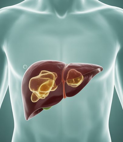
Fatty liver disease occurs when excess fat builds up inside liver cells, a process called hepatic steatosis. In 2023, international liver societies renamed the condition from non-alcoholic fatty liver disease (NAFLD) to metabolic dysfunction-associated steatotic liver disease (MASLD) to better reflect its metabolic origins.
The condition sits on a spectrum. Simple steatosis. There, fat is present but inflammation is minimal, can progress to steatohepatitis (MASH). There, active inflammation damages liver tissue. Left unchecked, MASH can lead to fibrosis, cirrhosis, and in rare cases liver failure.
According to the British Liver Trust, MASLD is now the most common liver condition in the UK. The NHS notes that most people with fatty liver disease experience no symptoms until the condition is advanced. This is precisely why early detection through body composition assessment is so valuable.
Why BMI Misses the Risk: The Role of Fat Distribution
Key Findings
BMI divides your weight by the square of your height. It cannot distinguish between muscle, bone, water, and fat. It cannot tell you whether your fat sits harmlessly under the skin on your arms and legs or clusters deep inside your abdominal cavity around vital organs.
Research published in the Journal of Clinical Medicine, analysing over 10,800 individuals using DEXA scanning, found that trunk fat percentage was the single strongest predictor of NAFLD in both obese and non-obese groups. People with a normal BMI but elevated trunk fat carried a significantly higher risk of fatty liver disease than those with higher overall weight but more peripheral fat storage.
This matters for a practical reason. If your GP measures only your weight and height, they may classify you as low risk while your liver is quietly accumulating fat. A DEXA scan provides the regional breakdown that BMI cannot.
How a DEXA Scan Detects Fatty Liver Risk
How DEXA Scanning Helps
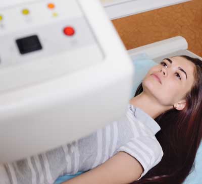
A DEXA (dual-energy X-ray absorptiometry) scan passes two low-dose X-ray beams through the body at different energy levels. The way tissues absorb these beams allows the scanner to separate fat mass, lean mass, and bone mineral content with high precision.
Critically for liver health, DEXA quantifies fat distribution by region. Your results report will show trunk fat mass and percentage, limb fat mass and percentage, the android-to-gynoid fat ratio (a comparison of abdominal versus hip and thigh fat), and estimated visceral adipose tissue volume.
Visceral fat is the metabolically active fat that surrounds organs in the abdominal cavity. Examples include the liver. Because visceral fat drains via the portal vein, the liver is directly exposed to high concentrations of free fatty acids released from this tissue. That portal delivery mechanism is one of the primary pathways through which visceral adiposity drives hepatic steatosis.
A study in BMC Gastroenterology found that NAFLD risk increased significantly when total body fat exceeded 32% in women and 27% in men, and when abdominal fat exceeded 21% in women and 14% in men. Your DEXA report places your numbers against exactly these thresholds.
The Android-to-Gynoid Ratio and Liver Health
How DEXA Scanning Helps
Your DEXA report includes a metric called the android-to-gynoid (A/G) fat ratio. The android region covers the abdomen and lower chest. The gynoid region covers the hips and upper thighs.
A higher A/G ratio indicates that your body stores proportionally more fat in the abdominal region. Multiple studies have linked an elevated A/G ratio to increased insulin resistance, dyslipidaemia, and hepatic steatosis. One analysis in the journal Obesity found that the A/G ratio outperformed both BMI and waist circumference as a predictor of metabolic dysfunction. Examples include fatty liver.
This ratio is particularly useful for people whose total body fat percentage appears moderate but whose distribution pattern leans heavily toward abdominal storage. These individuals, sometimes described as having normal-weight metabolic obesity, are the group most likely to be missed by conventional screening but flagged by DEXA.
Who Should Consider a DEXA Scan for Liver Health?
Understanding Liver Fat Risk
Not everyone needs a DEXA scan to assess liver risk. However, certain groups benefit significantly from the detailed fat distribution data it provides. You may want to consider a scan if you have been diagnosed with fatty liver on ultrasound and want to understand the underlying body composition driving it, if you have a family history of type 2 diabetes, metabolic syndrome, or liver disease, if your blood tests show elevated liver enzymes (ALT, AST, or GGT) without a clear explanation, if you carry most of your weight around your midsection despite a normal or near-normal BMI, or if you are taking medication for cholesterol, blood pressure, or blood sugar and want to track whether your body composition is improving alongside your blood markers.
A DEXA scan does not diagnose fatty liver disease directly. That requires imaging such as ultrasound or FibroScan, or in some cases a liver biopsy. What DEXA does is quantify the fat distribution pattern that predicts and drives the condition, giving you and your doctor actionable data for prevention or management.
Reducing Liver Fat Through Body Composition Change
Understanding Liver Fat Risk
The evidence on reversing early-stage fatty liver disease is encouraging. The European Association for the Study of the Liver (EASL) guidelines state that a sustained weight loss of 7 to 10 percent of body weight can resolve hepatic steatosis and even improve fibrosis in some cases.
However, total weight loss alone is not the full picture. Losing weight through crash dieting or excessive caloric restriction can reduce muscle mass alongside fat, potentially worsening your metabolic profile. The goal is targeted fat loss. In particular, visceral and trunk fat. While preserving or building lean tissue.
This is where serial DEXA scanning becomes a powerful monitoring tool. By comparing scans taken three to six months apart, you can track whether your intervention is reducing trunk fat and visceral adipose tissue specifically, not just lowering the number on the scales. A follow-up scan that shows reduced android fat with preserved lean mass confirms that your approach is working at the level that matters for liver health.
We explored this principle of fat loss versus muscle preservation in detail in our guide to intermittent fasting and body composition. This covers how different dietary strategies affect the ratio of fat to lean tissue lost.
Frequently Asked Questions
Understanding Liver Fat Risk
Can a DEXA scan detect fatty liver disease?
A DEXA scan does not image the liver directly. It measures the trunk fat and visceral adipose tissue levels that are the primary drivers of fatty liver disease, providing risk assessment data rather than a direct diagnosis.
I have a normal BMI but was told I have a fatty liver. How?
BMI measures total weight relative to height and cannot distinguish fat from muscle or determine where fat is stored. Up to 20% of people with fatty liver disease have a normal BMI but elevated trunk and visceral fat, a pattern DEXA scanning identifies clearly.
How much weight do I need to lose to reverse fatty liver?
Clinical guidelines from EASL suggest that a sustained loss of 7 to 10% of body weight can resolve hepatic steatosis. DEXA scanning helps confirm that the weight you are losing comes from fat. In particular, trunk and visceral fat, rather than muscle.
How often should I get a DEXA scan if I have fatty liver disease?
Most clinicians recommend a follow-up scan every three to six months when you are actively working to reduce liver fat through diet, exercise, or medical treatment. This interval allows enough time for measurable changes in body composition.
Is fatty liver disease serious?
Simple steatosis is reversible with lifestyle changes. If it progresses to steatohepatitis (MASH) and fibrosis, the consequences can be severe. Examples include cirrhosis and liver failure. Early detection and body composition monitoring significantly improve outcomes.
Book Your DEXA Body Composition Scan
Understanding Liver Fat Risk
If you have been diagnosed with fatty liver disease, suspect you may be at risk, or simply want to understand where your body stores fat and what that means for your metabolic health, a DEXA body composition scan provides the clinical-grade data you need.
DEXA London is based at 86 Harley Street, in the heart of London’s medical district. Scans take approximately 12 minutes and are reported by Dr Emil Gadimali. To book your scan or discuss whether DEXA is appropriate for your situation, call us on 0207 637 8227 or book online through our website.
Weight management next step
If your DEXA results reveal elevated trunk fat or visceral adiposity linked to fatty liver risk, a supervised medical weight-loss programme can help you achieve the 7 to 10% fat reduction that clinical guidelines recommend. CutKilo, the sister service to DEXA London, offers doctor-led Mounjaro treatment from Dr. Emil Gadimali. Start the CutKilo questionnaire to see if you are suitable.