


Rheumatoid Arthritis and Bone Density: Why a DEXA Scan Is Essential If You Have RA
Quick Answer
Rheumatoid arthritis significantly increases your risk of osteoporosis through two pathways: the disease itself drives inflammatory bone destruction, and the corticosteroids commonly used to manage flares accelerate bone loss further. A DEXA scan is the gold-standard method for measuring your bone density early and tracking changes over time, helping you and your doctor act before a fracture occurs.
If you have been diagnosed with rheumatoid arthritis, your joints are probably your primary concern. Pain, stiffness, and swelling demand daily attention. But beneath that visible inflammation, something less obvious may be happening: your bones could be losing density at a faster rate than expected.
Research published in Frontiers in Medicine shows that people with RA are roughly twice as likely to develop osteoporosis compared to the general population, and fracture risk rises accordingly. The good news is that early detection through a bone density DEXA scan allows you to intervene before a fracture happens. This guide explains why RA affects your bones, what your DEXA results mean in context, and what you can do to protect your skeleton while managing your condition.
How Rheumatoid Arthritis Affects Your Bones
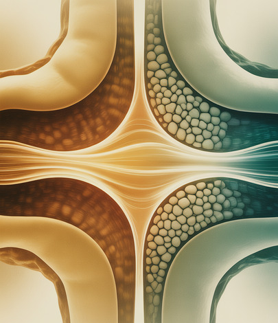
Rheumatoid arthritis is a systemic autoimmune condition. While it is best known for attacking the lining of your joints (the synovium), the inflammation it generates does not stay confined to those areas. Pro-inflammatory cytokines, particularly tumour necrosis factor alpha (TNF-α), interleukin-1 (IL-1), and interleukin-6 (IL-6), circulate throughout your body and directly influence bone metabolism.
These cytokines stimulate the production and activity of osteoclasts, the cells responsible for breaking down bone tissue. At the same time, they suppress osteoblasts, the cells that build new bone. The result is a net loss of bone density that occurs both locally, around inflamed joints, and systemically, across your entire skeleton. A peer-reviewed study in Rheumatology (Oxford Academic) confirmed that osteoclast overactivity is central to the generalised bone loss seen in RA patients, even in those who have never taken corticosteroids.
This systemic effect means that bone loss in RA is not limited to your hands and wrists. Your spine and hips are also at risk, and these are the sites where fractures carry the most serious consequences for your mobility and quality of life.
The Double Risk: Inflammation and Corticosteroid Treatment
Many people with RA face a compounded risk. The disease itself erodes bone through chronic inflammation, and one of the most commonly prescribed treatments for managing flares, corticosteroids such as prednisolone, has its own well-documented bone-thinning effect.
Corticosteroids reduce bone formation by suppressing osteoblast activity and simultaneously increase bone resorption by prolonging osteoclast survival. Even short courses of oral steroids, when repeated over months or years, can cause measurable bone loss. The National Institute for Health and Care Excellence (NICE) recommends that anyone expected to take oral corticosteroids for three months or longer should have their bone health assessed, and bone-protective treatment should be considered. For a more detailed look at how common medications affect skeletal health, our guide to medications that cause bone loss covers the broader picture.
The combination of inflammatory cytokine-driven destruction and steroid-induced suppression of bone formation creates what researchers describe as a “double hit” on the skeleton. This is why RA patients often lose bone density more rapidly than their peers, particularly during the first few years of active disease when inflammation is highest and steroid use is most frequent.
Who Should Get a DEXA Scan If They Have RA
Not every RA patient will develop osteoporosis, but the baseline risk is high enough that proactive screening makes clinical sense. The following groups should consider a DEXA scan sooner rather than later:
- Anyone with RA who is currently taking or has recently completed a course of oral corticosteroids lasting three months or more
- Postmenopausal women with RA, where the hormonal decline in oestrogen compounds the inflammatory bone loss
- Men over 50 with RA, particularly those with additional risk factors
- RA patients with low body weight (BMI under 19), a history of smoking, a family history of osteoporosis, or a previous fragility fracture
- Anyone with RA who has experienced height loss or a change in posture, which may indicate vertebral compression fractures
UK guidelines, including those from the National Osteoporosis Society and NICE, recognise RA as an independent clinical risk factor for osteoporosis. This means that RA patients are typically referred for DEXA screening earlier than the age thresholds recommended for the general population.
What Your DEXA Results Mean If You Have Rheumatoid Arthritis
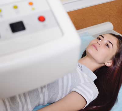
A DEXA scan measures bone mineral density (BMD) at key skeletal sites, typically the lumbar spine and hip. Your results are expressed as a T-score, which compares your bone density to that of a healthy 30-year-old adult. A T-score of -1.0 or above is considered normal. A score between -1.0 and -2.5 indicates osteopenia (reduced bone density), and a score of -2.5 or below indicates osteoporosis.
For RA patients, it is important to understand that fracture risk may be higher at the same T-score compared to someone without RA. The systemic inflammation associated with the disease affects bone quality as well as bone quantity, meaning that the structural integrity of the bone may be compromised even when the density measurement alone does not look alarming.
This is why the FRAX fracture risk assessment tool, recommended by the World Health Organisation, includes rheumatoid arthritis as a standalone risk factor. When your doctor enters your DEXA results into FRAX alongside your RA diagnosis, the calculated fracture probability will be higher than it would be for a non-RA patient with the same T-score. This adjusted risk estimate helps guide treatment decisions and determines whether bone-protective medication is warranted.
Protecting Your Bones While Managing RA
The encouraging reality is that bone loss in RA can be slowed, and in some cases partially reversed, with the right combination of disease management and bone-protective strategies.
Control the inflammation. The most effective way to protect your bones is to bring your RA under good control. Biologic therapies, particularly anti-TNF agents such as adalimumab and etanercept, have been shown in clinical trials to reduce not only joint inflammation but also systemic bone loss. By blocking TNF-α, these medications reduce the cytokine-driven osteoclast activity that erodes bone density. Research published in Nature Reviews Rheumatology confirms that early and sustained disease control with biologics is associated with better bone outcomes.
Optimise calcium and vitamin D. The NHS recommends that adults aim for 700 mg of calcium daily, with higher intakes sometimes advised for those at elevated risk of osteoporosis. Vitamin D is essential for calcium absorption, and deficiency is common in the UK, particularly during winter months. Your doctor may recommend supplementation, especially if your blood levels are low.
Stay physically active. Weight-bearing exercise, such as walking, stair climbing, and resistance training, stimulates bone formation and helps maintain density. RA can make high-impact activity uncomfortable, but lower-impact options like swimming, cycling, and physiotherapy-guided strength work still provide skeletal benefits. A physiotherapist familiar with inflammatory arthritis can help design a programme that strengthens bone without aggravating your joints.
Consider bone-protective medication. If your DEXA scan shows osteopenia or osteoporosis, your doctor may prescribe a bisphosphonate (such as alendronic acid) or denosumab. These medications reduce bone resorption and have been shown to lower fracture risk significantly. For RA patients already on corticosteroids, early initiation of bone-protective treatment is especially important.
How Often Should RA Patients Have a DEXA Scan
There is no single answer that applies to everyone, because the optimal scan frequency depends on your treatment, disease activity, and baseline bone density. However, the following general guidance applies:
- Baseline scan at diagnosis or when starting corticosteroids. If you have just been diagnosed with RA, or your rheumatologist is prescribing oral steroids for the first time, a DEXA scan establishes your starting bone density so that future changes can be tracked accurately.
- Follow-up every one to two years. For patients on ongoing corticosteroids or those with osteopenia at baseline, a repeat scan every 12 to 24 months allows your doctor to detect meaningful changes in bone density and adjust treatment accordingly.
- More frequent scanning if treatment changes. If you start or stop a biologic, change your steroid dose significantly, or begin bone-protective medication, a follow-up DEXA scan after 12 months can help evaluate the response.
- Less frequent scanning if stable. If your RA is well-controlled on a biologic, you are not taking steroids, and your most recent DEXA results were normal, scanning every two to three years may be sufficient.
Discuss the right schedule with your rheumatologist or GP. The key is not to wait until you fracture something before checking your bone density. Early detection gives you time to act.
Frequently Asked Questions
Does rheumatoid arthritis always cause osteoporosis?
No. Not everyone with RA will develop osteoporosis, but the risk is significantly higher than in the general population. Factors that increase the likelihood include long-term corticosteroid use, postmenopausal status, low body weight, and poorly controlled disease activity. A DEXA scan is the best way to find out where you stand.
Can biologic treatments protect my bones?
Yes, there is good evidence that biologic therapies, particularly anti-TNF agents, reduce systemic inflammation and slow the cytokine-driven bone loss associated with RA. Achieving and maintaining disease remission with biologics is one of the most effective bone-protective strategies available.
Should I get a DEXA scan if I have mild RA?
It depends on your overall risk profile. If you have mild RA and are not taking corticosteroids, your bone density may be unaffected. However, if you have additional risk factors, such as a family history of osteoporosis, low body weight, or smoking, a baseline DEXA scan is a sensible precaution.
What if my T-score is normal but I have RA?
A normal T-score is reassuring, but it does not mean your fracture risk is zero. Because RA affects bone quality as well as density, the FRAX tool adjusts your fracture probability upward when RA is entered as a risk factor. Your doctor can use the combined information to decide whether monitoring or preventive treatment is appropriate.
Can bone loss from RA be reversed?
Partial reversal is possible. Bringing inflammation under control with effective RA treatment, combined with bone-protective medications like bisphosphonates, has been shown to stabilise and in some cases improve bone density over time. The earlier the intervention, the better the outcome.
Book Your DEXA Bone Density Scan at Harley Street
If you have rheumatoid arthritis and have not yet had your bone density assessed, a DEXA scan gives you the objective data you need to make informed decisions about your skeletal health. At DEXA London, our Harley Street clinic offers consultant-reported bone density and body composition DEXA scans with results explained clearly on the day.
To book your scan, call us on 0207 637 8227 or use the booking form on our website. Whether you are looking for a baseline measurement or tracking changes over time, we are here to help you stay ahead of bone loss before it becomes a fracture.
Written by Dr Emil Gadimali